Cytology challenge: abdominal effusion in a 7-year-old Maine Coon cat.
Cells: 25,000/µl. Protein: 50 g/L.
What are your diagnoses? Because yes, there is more than one process to identify here, even if everything is related… Suspense…
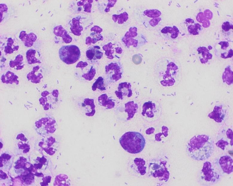
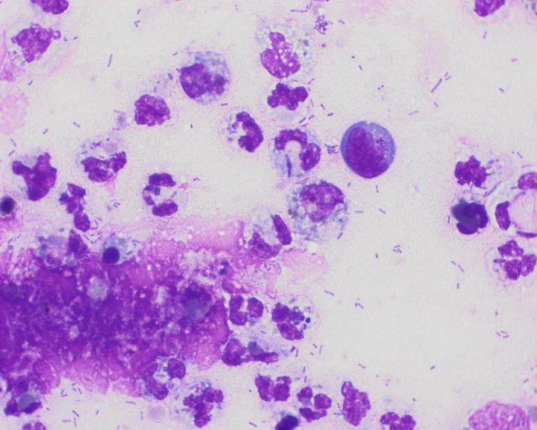
First thing: based on the numerical values, you are clearly dealing with an exudate caused by suppurative inflammation (with a predominance of neutrophils). The neutrophils are degenerated. Numerous bacteria are observed, both within the neutrophils and in the background of the smear. The inflammation therefore has a septic, bacterial component. We are dealing with septic peritonitis.
But is that all? The attentive observer will have noted the presence of occasional large, round cells with a high nuclear-to-cytoplasmic ratio: large lymphocytes, in other words. These lymphocytes consistently contain azurophilic granules in their cytoplasm. The monomorphism and large size of these lymphocytes suggest lymphoma.
Ultimately, this cat was suffering from a large-cell gastrointestinal lymphoma, which caused a loss of intestinal integrity with leakage of intestinal contents (including bacteria) and secondary septic peritonitis.